discontinued This product has been discontinued and is no longer available. You can still access this page for informational service purposes.
successor
This product's features have been integrated into a new model. Continue to successor

X線分析顕微鏡 XGT-7000V <販売終了しました>

真空チャンバ装備により軽元素の高感度分析が可能

用途:異物 分析、故障 解析、不具合 解析、RoHS 対応、スクリーニング、非破壊 分析、食の安全、鉛フリー、規制、鑑識、生態分析、生体分析

対象:プリント基板、実装基板、電子部品、家電、玩具、製薬、医薬品、食品、めっき

機能:マッピング、面分析、微小部 分析

事業セグメント: Scientific
製造会社: HORIBA, Ltd.

  • 10µm X線ガイドチューブ搭載
  • 革新のマッピングソフト スマートマップ搭載
  • デュアル バキューム チャンバ搭載

■デュアル バキューム チャンバ

  • 真空プローブ:含水、粉体など真空引きが困難なサンプルの軽元素分析を大気中で行えます。
  • 真空チャンバ:軽元素の感度をさらに向上。

充実のソフトウェア

元素マップを極めるスマートマップ
サンプルから得られる元素情報を全て保存。
一度スキャンしてしまえばサンプルがなくてもマップ像の追加や点・面分析も可能に。また、マッピングスピードも高速のため短時間分析を実現。

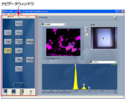
ナビゲータウィンドウにより操作手順をサポート
分析手順を知らなくてもナビゲータウィンドウによって分析手順がわかり、そのフロー(ボタン)を選択するだけで目的の操作ができます。各部分での操作は充実したアドバイスにより簡単にわかり、精度の良い自動定性と定性確認機能で、正確な分析が行なえます。
軽元素の感度向上

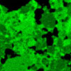
●隕石の分析例

Si
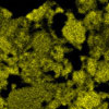
Mg
Mg

軽元素(Mg,Siなど)でも真空チャンバを使うことにより高感度な測定を実現。もちろん従来通り、大気中軽元素測定を可能とする真空プローブ機構も搭載しています。

XGT-7000Vは、さまざまな分野にフレキシブルに対応します。

薬剤
・錠剤内部、薬剤に混入した異物の分析等
・取り出し、断面出し等前処理なしで分析可能
自動車
・PM(粒子状物質)
・塗料、エンジンオイル中の異物等
・多点分析で複数位置も一度で測定
地質
・岩石、堆積物の分析
・風化状態の確認等
・分断しにくい大きな試料も
そのままセッティング可能
材料
・合金、金属、半導体、コンクリート等
・成形済みの各種成品の元素分析
バイオ
・植物や動物などの生体試料
・歯、臓器などの生体組織試料
・含水試料も分析可能
エレクトロニクス
・樹脂モールド中の不良解析
・ICパッケージの不良解析
・コンデンサ、LED等の不良解析
・非破壊で故障解析が可能
法科学
・塗膜片、インク成分の判定
・金属無機材、有機材の判定
・異同識別の手法
考古学
・陶磁器の染付け
・ガラス玉(トンボ玉)、勾玉
・絵画の顔料
・貴重な考古学試料の元素分析

測定原理

エネルギー分散型蛍光X線分析

測定元素

Na(ナトリウム)〜U(ウラン)

分解能

10 µm/100 µm

最大測定エリア

100×100 mm

設置寸法(本体)

670 (W)×820 (D)×760 (H) mm


Micro-XRF analysis for metal debris and particle analysis
Fast analysis of microscopic metal particles is an important application for engineering (e.g. engine wear analysis). Micro-XRF scanning and spot analysis of specific fragments identifies their chemical composition.
Micro-XRF for non destructive analysis of museum and archaeological objects
Pigments used in an ancient Nepalese manuscript have been analysed and assigned and an ancient glass burial ornament has been probed to identify the specific colouring additives used.
Biological applications of XRF microscopy
The effect of zinc on gastric ulcer healing has been investigated - mapped imaging of tissue samples shows evidence of zinc accumulation within the ulcerated tissue. In a separate study a fish otolith ('ear bone') has been analysed to reveal its heterogeneous elemental composition and physical structure.
Elemental micro-analysis of leaves using EDXRF
Calcium micro-nodules are identified within mulberry leaves, and XRF mapped imaging provides a correlation between nodule concentration and leaf age. In a seperate study, the uptake of heavy element pollutants by plants is investigated, with high resolution images quickly acquired to show the distribution of lead through the leaf.
Elemental analysis of single rice grains using XRF micro-analysis
Individual rice grains are analyzed by micro-XRF to examine the effect of grain polishing. The concentration of mineral elemnts found on a grain can be related to the degree of polish.
Micro-XRF analysis for the Electronics Industry
The combination of the XGT-5000’s ground breaking spatial resolution and sensitivity means it is the instrument of choice for fast analysis of electronic components, whether for analysis of restricted harmful elements (the WEEE/RoHS ‘lead free’ legislation), trouble shooting, or R&D.
Quality control and defect analysis in the electronics industry using micro-XRF
Troubleshooting and defect analyses of components embedded within opaque resins are described, on both individual components and complete circuit boards. Quantitative analysis to the ppm level is ideally suited for ensuring compliance to the WEEE/RoHS directives.
Fast thickness measurement of thin metal coatings by Micro-XRF
The penetrating nature of EDXRF analysis allows multi-layered samples to be characterised with a single measurement. With high spatial resolution even microscopic features such as bonding pads on circuit boards can be interrogated for composition and layer thickness.
Micro-XRF analysis for lead contamination in toys
A plastic toy is analysed for the presence of lead within its many components. Spot analysis shows concentrations of this harmful element can reach as high as 0.3%. XRF imaging allows its distribution across the toy to be quickly characterised.
Fingerprint imaging with micro-XRF
High spatial resolution elemental mapping experiments are demonstrated to provide a useful method of fingerprint analysis in situations where traditional methods struggle. Fingerprints on glossy paper and finely woven fabrics have been chemically treated, and subsequently imaged.
X-ray micro-analysis for pearl characterisation in forensic science
Simultaneous XRF and transmission x-ray imaging provides valuable insight into the composition and structure of pearls. Such information is vital for customs officials, who can quickly determine whether pearls are natural, cultured or imitation.
Gun Shot Residue analysis using X-ray fluorescence micro-analysis
Gunshot residue (GSR) consists of tiny particles expelled from a firearm when it is discharged. These particles, primarily composed of heavy metals, such as lead, barium, and antimony, can settle on the shooter and nearby surfaces, providing crucial forensic evidence in criminal investigations to determine if a firearm was recently used.

お問い合わせ

* 項目は必ずご記入ください。

HORIBAグループ企業情報